

DNA repair comprises cellular mechanisms that recognize and correct damage to genetic material. Damage arises during DNA copying and through ultraviolet light, ionizing radiation, chemicals and normal intracellular processes. It includes base loss, deamination, incorrect pairing and other changes.
The two DNA strands are complementary: information in an undamaged strand often enables repair of the other. However, they are not two identical copies, and not every lesion can be repaired without error. Different repair pathways address different types of damage.
Replication errors and proofreading
An incorrect nucleotide is sometimes incorporated during replication. Human replicative polymerases, particularly δ and ε, have 3′→5′ exonuclease proofreading activity that can remove an incorrect terminal nucleotide. Remaining errors may be corrected by mismatch repair (MMR). Polymerase β acts mainly in base excision repair, rather than serving as the principal replication polymerase. Error frequencies depend on the system and conditions; a universal sequence of 1:10⁶, 1:10⁷ and 1:10⁹ does not describe every cell.
Main stages of MMR
- Recognition of mispaired bases or small insertion/deletion loops.
- Selection of the strand to correct and removal of the error-containing segment.
- Synthesis of the correct sequence using the complementary template.
- Sealing the sugar–phosphate backbone by DNA ligase.
The classical Escherichia coli system
The following pathway describes methyl-directed MMR in E. coli, rather than all bacteria or humans. After replication, adenine in parental GATC sites is already methylated, while the new strand remains temporarily unmethylated. This difference helps direct correction to the new strand.
- MutS: recognizes the error. Bacterial MutS functions as a homodimer, a complex of two identical subunits. MutS₂ here denotes a dimer, not a separate MutS2 protein.
- MutL: coordinates interactions between the recognition complex and downstream enzymes.
- MutH: an endonuclease activated by the complex that nicks the unmethylated strand at a hemimethylated GATC site. This site may be some distance from the error.
- UvrD: DNA helicase II, which unwinds the duplex from the nick. It is not an exonuclease and does not itself excise the erroneous segment.
- Exonucleases: remove the new-strand segment containing the error. Enzyme choice depends on the direction of removal; examples include ExoI, ExoVII, ExoX and RecJ.
- DNA polymerase III and ligase: fill the gap and restore strand continuity; the single-stranded DNA-binding protein SSB also participates.
MMR in humans
Human mismatch recognition involves heterodimers: MutSα (MSH2–MSH6) and MutSβ (MSH2–MSH3), with different specificities. Subsequent factors include MutLα (MLH1–PMS2), EXO1, PCNA and other proteins. Correction of the new strand is linked to replication signals and strand discontinuities, rather than the bacterial GATC methylation/MutH scheme. Polymerase δ fills the gap and ligase seals the nick. This is a simplified description, not an exhaustive account of every pathway variant.
Base deamination
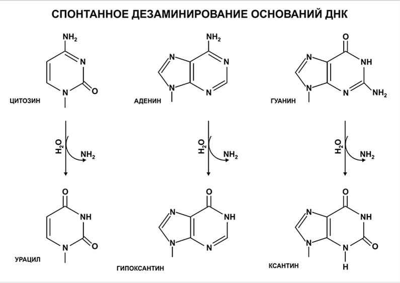
Spontaneous deamination is a common form of chemical DNA damage. Cytosine can become uracil, adenine hypoxanthine, and guanine xanthine. Appropriate repair systems recognize these altered bases. However, deamination of 5-methylcytosine produces thymine, a normal DNA base, so not every deamination product is foreign to DNA.
Base excision repair: BER
In typical human short-patch BER:
- A DNA glycosylase removes the damaged base, leaving an AP, or abasic, site.
- AP endonuclease APE1 cuts the sugar–phosphate backbone next to the AP site; it does not simply remove the entire deoxyribose residue.
- The lyase activity of polymerase β removes the deoxyribose phosphate residue, while its polymerase activity fills the gap with the correct nucleotide.
- DNA ligase seals the remaining nick.
Long-patch BER and other variants involve additional enzymes. Repair reduces the likelihood that changes become fixed but cannot guarantee correction of every lesion. DNA damage and an established genetic variant are not the same thing.
Sources and professional guidelines
- Paul Modrich: Mechanisms in E. coli and human mismatch repair
- ESRF: Structure of MutS binding to a G:T mismatch
- Primary study: Reconstitution of human mismatch repair
- Primary study: Human AP endonuclease and polymerase beta in base excision repair
Updated: 03.11.2026