

Hepcidin: A Regulator of Iron Homeostasis
Hepcidin and the anemia of chronic inflammatory diseases
Anemia is a hemoglobin concentration below the reference level appropriate to age and clinical state. It may accompany blood diseases and many other conditions. Erythrocyte counts and characteristics help identify the cause but do not necessarily change in parallel. Iron is required for hemoglobin and several enzymes of energy metabolism.
Iron deficiency can occur with or without anemia. Manifestations vary. In pregnancy, iron-deficiency anemia is associated with adverse maternal and perinatal outcomes; the cause and extent of deficiency require individual assessment.
Iron overload, including in hemochromatosis, can contribute to oxidative tissue injury. The liver, heart, and other organs may be affected. Not every elevated iron-status marker means hemochromatosis.
Both iron deficiency and iron excess adversely affect human health. To maintain iron within a relatively narrow permissible concentration range, absorption of exogenous iron and its metabolism must be tightly controlled.
Anemia of chronic inflammatory diseases (ACID) is an acquired complication of many chronic processes and is among the most prevalent forms of anemia of various origins. Its development is generally considered to result from disturbances at various stages of erythropoiesis, including:
- reduced sensitivity of precursor cells to erythropoietin (EPO);
- a shortened erythrocyte lifespan;
- inability of erythroblasts to utilize dietary and endogenous iron;
- low circulating iron due to its sequestration in tissue macrophages;
- impaired intestinal absorption of dietary iron.
Iron is essential for all organisms. It is a component of more than one hundred human proteins and enzymes with key roles in hematopoiesis, respiration, oxidation–reduction reactions, and various immune processes.
- An adult body contains approximately 4,000 mg of iron on average.
- Of the total iron content, 400–600 mg (10–15%) is incorporated into various heme-containing, transport, and other iron-containing proteins and enzymes.
- Circulating erythrocytes contain 2,600–2,800 mg (65–70%) of total body iron.
- Approximately 22 mg of iron is used daily for bone marrow erythropoiesis.
- Approximately the same amount (22 mg) is returned daily to the bloodstream from damaged erythrocytes processed by macrophages of the reticuloendothelial system.
- About 800 mg (20%) is stored in tissue macrophages.
- Normally, approximately 1–2 mg of iron is lost from the human body daily. This is replaced by exogenous dietary iron absorbed by duodenal enterocytes and released into the bloodstream as needed.
Maintaining and redistributing iron between spatially separated compartments is a tightly controlled dynamic process regulated according to the body's requirements.
Discoveries of proteins involved in iron absorption, storage, and reutilization have substantially expanded understanding of its metabolism. Hepcidin is a key hormonal regulator of iron availability.
Mature hepcidin consists of 25 amino acid residues and contains four disulfide bonds. The liver synthesizes an 84-amino-acid prepropeptide that undergoes further processing. Prohepcidin and mature hepcidin-25 are different analytical targets.
Hepatocyte hepcidin production decreases in response to stimulation of iron export from cells into the bloodstream when serum iron is low or erythropoiesis is increased. Conversely, hepcidin production rises when iron export from reticuloendothelial cells into the systemic circulation needs to be reduced because of excess serum iron or inflammatory processes.
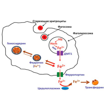
Ferroportin exports iron from enterocytes and cells that recycle or store it. In blood, iron binds transferrin and is delivered, among other sites, to bone marrow for erythropoiesis. Stores are mainly held in ferritin. Transport depends on hepcidin regulation of ferroportin as well as unsaturated transferrin.
Iron is stored by ferritin, a protein found predominantly in macrophages of the liver, spleen, and bone marrow. Ferritin*** is the main iron-storage protein and contains approximately 20% of the body's iron.
* Ferroportin is a membrane iron exporter present, among other sites, on the basolateral enterocyte surface and on macrophages. It transports Fe2+; ferroxidases including hephaestin and ceruloplasmin provide oxidation for transferrin binding. Hepcidin binds ferroportin, restricting iron export.
** Transferrin is a transport glycoprotein of approximately 80 kDa with two Fe3+-binding sites. The iron-free protein is called apotransferrin. Transferrin delivers iron to cells through appropriate surface receptors.
*** Ferritin is a complex of iron ions with apoferritin. The latter is a protein of approximately 450 kDa composed of 24 subunits forming a cavity capable of binding up to 4,000 iron ions. Normally, each ferritin molecule contains approximately 2,200 iron ions on average.
Ferritin primarily stores iron within cells. Serum ferritin is used as a marker of stores, taking inflammation and other influences into account; it is not the main iron transport protein in blood.
The connection between hepcidin and iron homeostasis was first demonstrated by Pigeon and colleagues, who showed that excess serum iron leads to increased expression of the hepcidin precursor in the liver. They recorded a similar effect when simulating inflammation by injecting mice with lipopolysaccharides (LPS).
Experiments in transgenic mouse lines established that hepcidin negatively regulates iron uptake in the small intestine and its release from macrophages. This conclusion was based on the development of hemochromatosis in a line of knockout mice (mice lacking the hepcidin gene (HAMP)). Conversely, studies in transgenic mice with increased hepcidin gene (HAMP) expression showed that increased hepcidin production causes acute iron deficiency and, consequently, death shortly after birth. These findings indicate that hepcidin blocks not only intestinal iron absorption and macrophage iron release but also placental transport. Further studies established its inhibitory effect on iron transport throughout the body, including different cell types.
Hepcidin often rises during inflammation, particularly through IL-6, and helps restrict iron availability for erythropoiesis. However, its level also reflects iron stores, hematopoietic activity, and accompanying conditions, so it is not necessarily elevated in every inflammatory state.
Experiments involving volunteers showed that, in lipopolysaccharide-induced inflammation, serum IL-6 rises after three hours, while peak hepcidin expression and a sharp decline in serum iron are observed by six hours. Similar findings were obtained in inflammation initiated by bacterial infections and in tumor growth. In both cases, anemia developed alongside increased serum IL-6 and hepcidin concentrations.
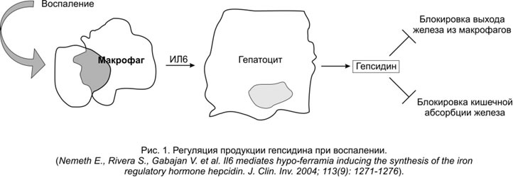
Figure 1 presents the regulation of hepcidin production during inflammation induced by pathogen-specific macromolecules (particularly LPS), infectious agents, transformed tumor tissues, and other factors. According to this model (Nemeth and colleagues), inflammation induces increased macrophage production of interleukin-6, which in turn activates hepatocyte hepcidin production. The resulting excess hepcidin, whose level may rise hundreds of times, blocks iron release from macrophages and intestinal iron absorption, thereby causing hypoferremia.
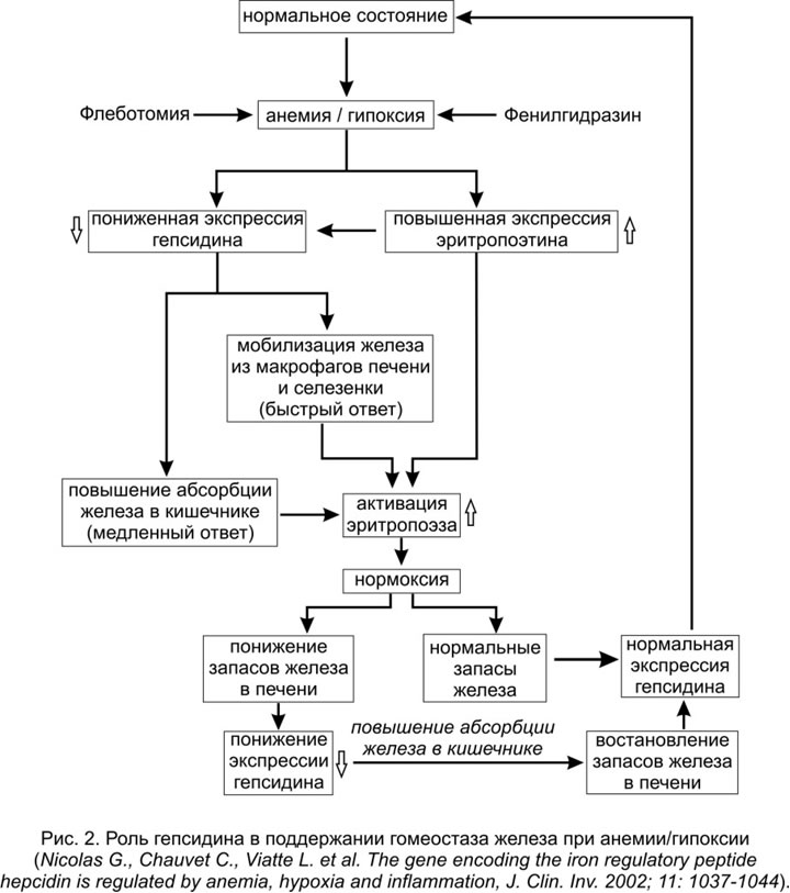
Anemia and increased erythropoietic demand usually suppress hepcidin and increase iron availability. Hypoxia activates intracellular HIF signaling and renal erythropoietin production; HIF should not be described as an ordinary serum regulator. The overall effect depends on the combination of these signals with inflammation and iron stores (Fig. 2).
These data support the conclusion that hepcidin is the principal iron-regulatory hormone and a key mediator of ACID.
Other evidence of hepcidin's key role in iron metabolism includes:
- anemia resistant to iron treatment in patients with hepcidin-producing tumors;
- hereditary hemochromatosis caused by mutations in the hepcidin gene (HAMP).
Subnormal dietary iron absorption and delayed macrophage iron release are clearly evident in infections, tumors, and chronic inflammatory diseases. In these conditions, iron uptake by erythropoietic precursor cells has been shown to be restricted even when iron stores are sufficient in the early stages of anemia.
In addition to regulating iron absorption, hepcidin can disrupt bacterial membranes, creating an unfavorable microenvironment for microorganisms entering the bloodstream.
Restricting iron availability contributes to defense against some microorganisms. Its effect depends on the organism and conditions; it cannot be reduced to obligatory suppression of one iron-containing enzyme in every microorganism.
Hepcidin and ACID in Rheumatoid Arthritis
Rheumatoid arthritis (RA) is a chronic inflammatory disease that can affect joints and other organs. It has substantial medical and social consequences.
Anemia frequently accompanies RA. Its prevalence depends on the population studied and disease activity. Anemia of inflammation, true iron deficiency, and combinations can occur.
Research has confirmed the important role of the hepcidin–ferroportin axis in anemia of inflammation. It regulates intestinal iron entry and distribution between cells and blood.
Hepcidin is an oligopeptide consisting of 25 amino acid residues and four disulfide bridges. It is synthesized in hepatocytes as a precursor of 84 amino acid residues.
The link between inflammation and hepatocyte hepcidin production is mediated by interleukin-6 (IL-6), whose macrophage production is induced by inflammation (Fig. 1).
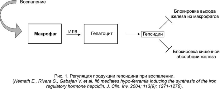
Table 1 presents the clinical and laboratory characteristics of patients with RA according to the type of anemia.
Table 1. Blood hemoglobin and serum hepcidin and IL-6 levels and the IL-6/hepcidin ratio in the following groups of patients with RA: 1) without anemia, N=29, control group; 2) with confirmed iron-deficiency anemia (IDA), N=17; 3) with confirmed ACID without IDA, N=13.

* Serum concentration ratios normalized to 1 (control group).
These data show a direct correlation between serum IL-6 and hepcidin concentrations in ACID, consistent with the model of hepcidin regulation during inflammation shown in Fig. 1. Conversely, patients with RA and confirmed IDA have lower serum hepcidin than the control group, promoting dietary iron absorption from the intestine and iron release from macrophages.
Hepcidin can complement the differential diagnosis of anemia of inflammation and iron-deficiency anemia, but one result does not replace the complete blood count, ferritin, transferrin saturation, inflammatory markers, and clinical assessment. Mixed anemia is possible.
Treatment depends on the cause of anemia and confirmed iron deficiency. Iron medication should not automatically be prescribed or excluded on hepcidin alone. Different measurement methods require their own reference intervals.
Tocilizumab is an IL-6 receptor antibody used for particular RA treatment indications; it is not a direct hepcidin antagonist or a universal treatment for any anemia. IL-6 suppression may lower hepcidin and improve inflammatory anemia. A rheumatologist prescribes treatment with regard to contraindications and infection risk.
Research discusses links between inflammation, iron accumulation, and oxidative processes in synovial tissues. These mechanisms alone do not establish indications for a particular treatment.
References
Andrews N. C. Anemia of inflammation: the cytokine-hepcidin link. J Clin Invest 2004;113:1251-3.
Demirag M., Haznedaroglu S. R., Banu S. K. et al. Circulating hepcidin in the crossroads of anemia and inflammation associated eith rheumatoid arthrits. Inter Med 2009;48:421-6.
Demirjian S. G., Nurko S. Anemia of chronic kidney disease: when normaly becomes undesirable. Cleve Clin J Med 2008;75(5):353-6.
Ganz T. Hepcidin in iron metabolism. Curr Opin Hematol 2004;11(4):251-4.
Genovese M. C., McKary J. D., Nasonov E. L. et al. Interleukin-6 receptor inhibition with tocilizumab reduced disease — modifying antirheumatic drugs: the tocilizumab in combination with traditional disease — modifying antirheumatoic brug therapy study. Arthr Rheum 2008;58(10):2968-80.
Jayaranee S., Sthaneshwar P., Sokkalingam S. Serum prohepcidin concentrations in rheumatiod arthrits. Pathology 2009;41(2):178-82.
Jaworski J., Maslinski W., Pazdur J. et al. Decreased expression of integrins by hematopoietic cells in patients with rheumatoid arthrits and anemia: relationship with bone marrow cytokine levels. J Investing Allergol Clin Immunol 2008;18(1):17-21.
Kim H. R., Kim K. V., Yoon S. Y. et al. Serum pro-hepcidin could reflect disease activity in patients with rheumatoid arthritis. J Korean Med Sci 2010;25(3):348-52.
Masson C. Rheumatoid anemia. Joint Bone Spine 2011;78(2):131-7.
Nemeth E., Valore E. V., Territo M. et al. Hepcidin, a putative mediator of anemia of inflammation, is a type II acutephase protein. Blood 2003;101:2461-3.
Nemeth E., Rivera S., Gabayan V. et al. IL-6 mediates typoferremia of inflammation by inducting the synthesis of the iron regulatory hormone hepcidin. J Clin Invest 2004;113:1271-6.
Nicolas G., Chauvet C., Viatte I. et al. The gene encoding the iron regulatory peptide hepcidin is regulated by anemia, hypoxia and inflammation. J Clin Invest 2002;(110):1037-44.
Nikolaisen C., Figenschau Y., Nossent J.C. Anemia in early rheumatoid arthritis is associated with iterleukin 6-mediated bone marrow suppression, but has no effect on disease course or mortality. J Rheumatol 2008;35(3):380-6.
Nissenson A. R., Goodnough I. T., Dubois R. W. Anemia: not just an innocent bystander? Arch Intern Med 2003;163:1400-4.
Pigeon C., Ilyin G., Courselaud B. et al. A new mouse liver-specific gene, encoding a protein homologous to human antimicrobial peptide hepcidin, is overexpressed during iron overload. J Biol Chem 2001;7811-9.
Weiss G., Goodnough I. T. Anemia of chronic disease. New Eng J Med 2005; 352(10):1011-23.
Galushko E. A. Anemia of chronic disease. Nauch-praktich revmatol 2009;3(Suppl.):70-8.
Galushko E. A., Belenky D. A., Aleksandrova E. N., Kashnikova L. N. Nauch-praktich revmatol 2012;52(3):19-24.
Levina A. A., Kazyukova T. V., Tsvetaeva N. V. et al. Hepcidin as a regulator of iron homeostasis. Pediatriya 2008;8(1):67-75.
Litvinov A. V., Ivanyan A. N., Nikiforovich I. I. Rossiysky vestnik akushera-ginekologa 2010; 1: 11-14.
Nasonov E. L., Panasyuk E. Yu., Buldakov S. G. et al. Efficacy and safety of tocilizumab in rheumatoid arthritis (interim results of a Russian multicenter study). Nauch-praktich revmatol 2010;2:21-30.
The historical laboratory announcement reported introduction of hepcidin testing on April 1, 2015 using HEPCIDIN-25(Bioactive) ELISA. Current availability and method characteristics should be confirmed with the laboratory.
The historical European population values below relate to the specific IBL Ref.54161 assay; they are not universal diagnostic thresholds.

Historical Diagen Plus data for the Armenian group studied, with hemoglobin of 12.9±1.1 g/dL, are retained below. They do not establish a universal population reference interval.

Sources and professional guidelines
- Nemeth et al.: Hepcidin Regulation of Ferroportin, 2004
- Structure/Activity Relationships of Hepcidin
- Primary Research: Human Serum Transferrin Structure and Iron Binding
- Hepcidin Assay Standardization Study
- FDA: Actemra Prescribing Information
Updated: 03.11.2026