

Last updated
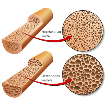
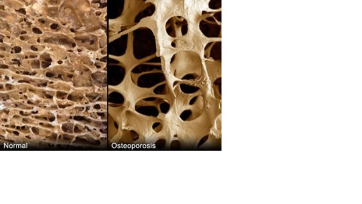
Osteoporosis is a skeletal disease in which bone strength and resistance to fracture are reduced. Both bone mineral density and bone quality matter. The main purpose of assessment and treatment is to evaluate and reduce fracture risk, rather than simply change the result of an individual laboratory test.
How bone tissue is renewed
Bone is constantly being remodelled: osteoclasts remove areas of old tissue, while osteoblasts form new tissue. These coordinated processes are called bone remodelling. When resorption exceeds formation over a prolonged period, bone mass decreases and its microarchitecture deteriorates. Type I collagen forms the organic framework, while mineral components provide rigidity. A change in an individual metabolic pathway does not explain every case of osteoporosis.
Risk factors and secondary causes
The likelihood of osteoporosis and fractures increases with age. After menopause, falling estrogen levels can accelerate bone loss. Men are also affected. Relevant factors include a previous low-trauma fracture, family history, low body weight, smoking, excessive alcohol intake, insufficient physical activity and falls.
Secondary bone loss may be associated with hypogonadism, hyperthyroidism, hyperparathyroidism, glucocorticoid excess, malabsorption, some kidney diseases and other conditions. Long-term glucocorticoid use and certain cancer treatments require a specific assessment of fracture risk. In diabetes, fracture risk may be increased even when bone mineral density is normal or relatively high: bone density alone is therefore insufficient to rule out an increased fracture risk.
Not every bone disorder is osteoporosis. Osteomalacia, metastatic disease, kidney-related mineral disorders and Paget disease require separate evaluation. Paget disease affects remodelling in particular skeletal sites; it should not be treated as simply another form of generalized low bone density or attributed to a proven viral cause.
How the diagnosis is established
The clinician considers the circumstances of fractures, height, posture, medications, other medical conditions and imaging findings. The principal method for measuring bone mineral density is dual-energy X-ray absorptiometry (DXA), usually at the lumbar spine and proximal femur. Suspected vertebral fractures may require radiographs or vertebral fracture assessment using DXA.
- In postmenopausal women and men aged 50 or older, a T-score ≤ −2.5 at an appropriate measurement site is a densitometric diagnostic criterion. The result is interpreted alongside the clinical findings.
- In premenopausal women and men younger than 50, Z-scores are generally used. A value ≤ −2.0 indicates bone density below the expected range for age, but does not by itself establish a diagnosis of osteoporosis.
- A low-trauma fracture can have important diagnostic implications even when the T-score is above −2.5. Risk assessment extends beyond the threshold of an individual measurement.
Peripheral bone ultrasound and ordinary radiography do not replace central DXA for standard densitometric diagnosis. FRAX helps estimate the probability of fracture over 10 years using clinical factors and, when available, femoral-neck bone density. The model and intervention thresholds depend on the country and applicable clinical guidance. FRAX is not designed to assess the short-term response to treatment.
Why laboratory investigations are useful
Laboratory investigations help identify secondary causes of bone loss and assess mineral metabolism and treatment safety. Depending on the clinical situation, the clinician may request a complete blood count, albumin-adjusted or ionized calcium, phosphate, alkaline phosphatase, creatinine and kidney-function assessment, and 25-OH vitamin D. Parathyroid hormone, TSH, sex hormones, coeliac testing and other investigations are added when indicated. The same extended laboratory panel is not necessary for every patient.
β-CTX: a marker of resorption
Breakdown of type I collagen releases fragments, including C-terminal telopeptides. Measuring blood β-CTX helps assess the rate of bone resorption and the biochemical response to antiresorptive treatment. A high or low β-CTX result does not independently diagnose or exclude osteoporosis.
Results are affected by time of day, food intake, kidney function, a recent fracture and other factors. To obtain comparable measurements, β-CTX is generally measured in the morning after an overnight fast, following the laboratory's instructions. Follow-up samples should be taken under similar conditions and preferably analysed using the same method. Changes are considered together with treatment, adherence and clinical information.
P1NP: a marker of bone formation
The N-terminal propeptide of type I procollagen (P1NP) is released during collagen formation and is used as a marker of bone formation. P1NP and β-CTX have among the most standardized applications of the bone turnover markers. They may help monitor treatment but do not replace DXA, fracture assessment or investigation of the cause. Total and intact P1NP are measured by different assays; the characteristics of the method are particularly important in impaired kidney function.
Osteocalcin and N-MID osteocalcin
Osteocalcin is a vitamin K-dependent protein produced mainly by osteoblasts. Both intact protein and fragments can be measured in blood; N-MID assays measure the corresponding forms. Osteocalcin reflects bone turnover but is affected by the assay, kidney function and other factors. Its level alone cannot reliably predict osteoporosis, establish rickets or determine the effectiveness of every form of treatment.
Parathyroid hormone (PTH)
PTH is produced by the parathyroid glands and helps regulate calcium and phosphate metabolism through effects on bone and the kidneys and on the formation of active vitamin D. Measurement is indicated in selected investigations of abnormal calcium and certain conditions associated with bone loss. Results are interpreted with calcium, vitamin D and kidney function. Sustained PTH excess and intermittent therapeutic administration of drugs acting through its receptor have different effects; it is incorrect to describe all PTH activity as simply suppressing osteoblasts.
25-OH vitamin D
Vitamin D status is generally assessed by measuring 25-hydroxyvitamin D, or 25(OH)D. Vitamin D contributes to calcium absorption and bone mineralization. Severe deficiency can cause osteomalacia, which must be distinguished from osteoporosis. A normal 25(OH)D level does not exclude osteoporosis, and a low result does not automatically explain every fracture. Measuring the active metabolite 1,25(OH)₂D is not a routine substitute for 25(OH)D when assessing vitamin D status.
The need for testing and correction of deficiency depends on the patient's condition, diet, medications and intestinal, liver and kidney diseases. The formulation and dose are chosen individually. Taking high doses of vitamin D or calcium without medical guidance can be unsafe.
Calcitonin
Calcitonin is produced by thyroid C-cells and participates in the regulation of bone turnover. Its concentration is not a standard laboratory criterion for diagnosing or monitoring osteoporosis. A low calcitonin level after menopause is not an established independent cause of the disease. Calcitonin testing has other indications, including the investigation of certain thyroid disorders.
What genetic tests can and cannot show
Heredity influences skeletal characteristics, but common age-related osteoporosis is multifactorial. Findings from studies of individual genetic associations cannot automatically be translated into personal fracture predictions or medication choices. A common polymorphism is not equivalent to a pathogenic variant causing a single-gene disorder and should not, without justification, be described as a dominantly inherited osteoporosis mutation.
COL1A1
COL1A1 encodes the α1 chain of type I collagen. Rare pathogenic variants in COL1A1 and COL1A2 can cause osteogenesis imperfecta and other connective-tissue disorders. This is different from testing a common Sp1-binding-site variant, rs1800012. Associations with bone density and fractures have been investigated, but an individual genotype does not establish a diagnosis or determine the need for surgery, supplements or medication. A genetic report should identify the variant precisely and specify the reference sequence.
VDR
VDR encodes the vitamin D receptor, which regulates the expression of genes involved in processes including calcium metabolism. BsmI is a common variant that has been studied. Its presence does not mean that vitamin D deficiency, a fracture or a particular treatment response is inevitable. Results should use a precise variant identifier and reference sequence. Standard osteoporosis management is not based on this polymorphism alone.
LCT and the MCM6 regulatory region
LCT encodes lactase, the enzyme that breaks down lactose. Variants in the MCM6 regulatory region may influence persistence of lactase activity into adulthood. The −13910C>T marker has mainly been studied in certain populations and does not describe every cause of lactose intolerance. Congenital lactase deficiency, an age-related decline in lactase activity and symptoms after consuming dairy products are distinct concepts.
A genetic result alone does not determine whether symptoms are present and does not diagnose osteoporosis. If someone limits dairy products, calcium and protein intake from other suitable sources should be assessed. Nutrition is adjusted to individual tolerance and requirements, rather than imposing identical restrictions on everyone with a particular genotype.
FDPS
FDPS encodes farnesyl diphosphate synthase, an enzyme in the mevalonate pathway. It is involved in the mechanism through which nitrogen-containing bisphosphonates affect osteoclasts. Possible associations between individual FDPS polymorphisms, bone density and treatment response have been investigated, but this testing is not a standard way to choose a bisphosphonate or its dose. An individual variant cannot predict a fixed amount of bone loss.
ESR1
ESR1 encodes estrogen receptor α. Estrogen signalling is important for skeletal maintenance, and common ESR1 variants have been investigated in bone-metabolism research. These variants do not independently establish the cause of osteoporosis, infertility or pregnancy loss. Decisions about menopausal hormone therapy should not be based on an individual genetic marker.
Testing for rare inherited bone disorders may be appropriate in unusually early or recurrent fractures, characteristic clinical features and a relevant family history. A specialist selects the investigations after clinical assessment. A panel of common COL1A1/VDR/FDPS/ESR1 polymorphisms does not replace this evaluation and is not standard osteoporosis screening.
Treatment and follow-up
The treatment plan considers fracture risk, secondary causes, kidney function, other medical conditions and the patient's preferences. Appropriate physical activity, strength and balance exercises, fall prevention, adequate nutrition and correction of confirmed calcium or vitamin D deficiency are important. These measures do not always replace medication in a person at high fracture risk.
Depending on the clinical situation, options include antiresorptive drugs such as bisphosphonates and denosumab, and bone-anabolic treatment for selected indications. Menopausal hormone therapy is considered individually in the light of its benefits and risks. Hormones plus bisphosphonates should not be presented as a universal regimen, and treatment should not be selected from the results of a common-polymorphism panel.
For denosumab, advance planning of treatment duration and subsequent therapy is particularly important. Discontinuation or a substantial delay without discussion with the clinician can lead to a rapid increase in bone resorption and a higher risk of vertebral fractures. Prescribed medication should not be changed or stopped independently.
Follow-up considers new fractures, changes in height and symptoms, adherence, adverse effects and, when indicated, repeat DXA and turnover markers. The interval between DXA scans is individualized. Measurements should preferably use comparable equipment and account for precision error and the least significant change. A small numerical difference does not always represent real bone loss. A change in β-CTX or P1NP helps assess the expected biochemical effect of a particular drug, but does not by itself quantify the reduction in fracture risk.
Sources and professional guidelines
- ISCD — Official Adult Positions (2023)
- NOGG — Fracture risk assessment and case finding
- NOGG — Pharmacological treatment options
- NOGG — Management and monitoring of osteoporosis
- NIAMS — Osteoporosis: diagnosis and treatment
- MedlinePlus — Vitamin D test
- MedlinePlus — Parathyroid hormone test
- MedlinePlus — Calcitonin test
- MedlinePlus Genetics — COL1A1
- MedlinePlus Genetics — VDR
- MedlinePlus Genetics — Lactose intolerance
- ClinVar — COL1A1 Sp1-binding-site variant
- Primary study — Farnesyl diphosphate synthase as a bisphosphonate target
Updated: 03.11.2026